Cardiac Procedures

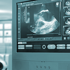
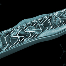
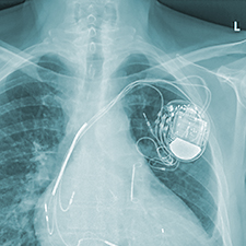
Depending on the patient’s condition and diagnosis, we may recommend a particular treatment or cardiac procedure, such as those outlined below. Urgent angiography and stenting, cardiac imaging and cardiac pacing are performed in University Hospital Waterford and UPMC Whitfield.

Active symptoms that might suggest cardiac disease include chest pain or discomfort, breathlessness, and palpitations. We provide urgent review and complete investigation from basic consultation through to invasive evaluation and treatment.

Transoesophageal Echo (TOE)

This ultrasound examination involves the patient swallowing the echo probe into the oesophagus (food pipe). This allows the cardiologist to acquire detailed images of the cardiac anatomy, especially the heart valves. This test is often used to assess and plan for heart valve surgery. It is performed under sedation.

See Video

Dobutamine Stress Echo

This is the chemical equivalent of having an exercise stress test performed during an echocardiogram. A chemical called Dobutamine is administered through a vein. This causes the heart to beat faster, and we can directly see how the heart performs under pressure. We can also identify areas of the heart that may be damaged, performing poorly or not receiving an adequate blood supply.

CT Calcium Scoring

This test is a special x-ray using computed tomography (CT). The test measures calcium build-up in the walls of the coronary arteries. Since calcium is often deposited in heart arteries that are narrowed, and can readily be seen on CT scanning, this test can be used to look for the early signs of heart disease.

Coronary Angiogram

A coronary angiogram is a diagnostic test to visualise your coronary arteries. The procedure is performed by Dr Owens in the cardiac catheterisation lab using specialised X-ray equipment. Coronary angiography is carried out using local anaesthetic while the patient is awake. The test usually lasts around 10-15 minutes.

Generally, the cardiologist uses a blood vessel in your right wrist (radial artery) and occasionally the left wrist to perform the examination. In some patients, it is not possible to use the arteries in either wrist. In such instances, the angiogram is performed using the artery at the top of your leg.

An angiogram is performed to confirm and define the extent of coronary artery disease (blockages in your heart arteries). The test is performed in patients with risk factors for heart disease, those presenting with symptoms for heart disease and in patients with abnormal non-invasive tests (e.g. exercise stress test or CT calcium scores).

At the end of the examination, the cardiologist will explain the results of the angiogram and recommend a treatment plan. There is a period of observation by the nursing staff for 2-3 hours after the test in the cardiac day ward. This is to ensure there are no post procedural complications. You will not be able to drive home after the angiogram. You can resume normal activities the following day.

See Video

Cardiac Stenting

Cardiac stents are tiny metallic scaffolds that are positioned inside the coronary artery using X-ray guidance. The purpose of the stent is to provide permanent scaffolding to keep the coronary artery open at a site where the artery has become narrowed or blocked with atheroma; atheroma is fatty tissue deposited in the arteries of people with heart disease. More than one stent may be required to successfully complete treatment of the blocked coronary artery. Stents may be placed in more than one of your coronary arteries during the procedure.

Most modern stents are referred to as drug eluting (coated) stents. This term indicates that the stent is covered externally by a drug that is released into the wall of your coronary artery over the next 3 months after the procedure. The rationale for using drug eluting stents is to minimise the risk of recurrence of the blockage in your coronary artery.

Following successful treatment of your coronary blockages with stenting, you will be observed overnight in a specialised cardiology ward. You will be reviewed the following morning by the cardiology team and will likely be discharged home. Following this procedure you will be seen in the office for an appointment 6-8 weeks after the stenting procedure to review your medications and discuss future management of your heart disease.

See Video

Pacemaker Implantation

A pacemaker is a small electrical device which is used to treat some abnormal heart rhythms that can cause your heart to beat too slowly or miss beats. Having a pacemaker can greatly improve your quality of life and for some people can be lifesaving. They’re smaller than an average matchbox and weigh about 20 to 50 grams. They sit under the skin, just below your collar bone, and have one or more leads which are placed into your heart through a vein.

Pacemakers are fitted under a local anaesthetic, with sedation if requested, so you may feel very sleepy. After the pacemaker is fitted, you’ll usually stay overnight in hospital and your pacemaker will be checked thoroughly before you leave. It’s normal to feel slightly sore for a few days afterwards, but most people find that they can get back to their normal lifestyle fairly quickly. You’re not allowed to drive a car for a week after your pacemaker is fitted (regulations differ for occupational driving).

See Video

Defibrillator Implantation

An implantable cardioverter defibrillator (ICD) is a small device which can treat people with dangerously abnormal heart rhythms. Abnormal heart rhythms (or arrhythmias) can cause your heart to beat too quickly, too slowly or in an irregular pattern. The defibrillator is inserted just under your collar bone. It looks similar to a pacemaker and is a little bigger than a matchbox. It is made up of a pulse generator (battery powered electronic circuit) and one or more electrode leads which are placed into your heart through a vein.

An implantable defibrillator is inserted under local anaesthetic, with sedation if required, so you may feel very sleepy. It takes about half an hour to implant an ICD, the time it takes will depend on the type of device you’re having. You will usually need to stay overnight in hospital and your ICD will be checked electronically before you leave.

See Video

Cardiac Resynchronisation Therapy

Cardiac resynchronisation therapy (CRT) is used to help improve the quality of the heart’s contractions, and the symptoms associated with heart failure. The procedure involves implanting a pacemaker usually just below the collarbone. Three wires (leads) connected to the device monitor the heart rate, and make the heart beat in a more co-ordinated way.

Loop Recorder Implantation

An implantable device called a loop recorder may be recommended to patients with a diagnosis of syncope (unexplained loss of consciousness). This small device allows for remote monitoring of your heart rhythm and helps to diagnose the cause of collapses, blackouts and dangerous heart rhythms.

This is a day case procedure carried out in the catheterisation lab. The device is usually implanted under local anaesthetic under the skin just to the left of the breast bone; this is a very small device, and is ‘injected’ through a tiny skin puncture. The procedure only takes 5 minutes to complete. The patient will be fully reviewed by a member of the cardiology team prior to discharge to explain the procedures used to monitor the data from the loop recorder. The long battery life means the loop recorder stays in place for up to 2-3 years, but it’s monitoring life is sometimes as long as four years.

There is a period of observation by the nursing staff for 2-3 hours after the procedure in the cardiac day ward. This is to ensure there are no post procedural complications.

See Video

Cardioversion

A cardioversion may be recommended when a patient has an abnormal heart rhythm. This day case procedure is performed in the Coronary Catheterisation Laboratory or General Theatre, using sedation administered by an anaesthetist. While the patient is sedated, an electrical shock is delivered to the heart to reset the heart’s electrical rhythm. Following the cardioversion, there is a period of observation. Please note that after this procedure, you will not be permitted to drive home.

See Video